Research Involving Animals – Division of Biomedical Services
Preclinical imaging facility
 The preclinical imaging facility is located behind the specific pathogen free (SPF) barrier at the preclinical research facility. It consists of state-of the-art, in-vivo imaging systems.
Our imaging systems
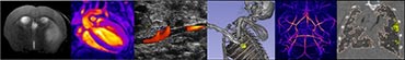
Micro-CT
The Quantum-FX X-Ray micro computed tomography (CT) scanner uses low dose X-rays (13-300 mGy) to provide high-resolution 3D images of bone structure and surrounding soft tissue. Whole body data can be acquired in less than 1 minute. Respiratory gating software can be used to reduce breathing motion-related artefacts. CT images can be co-localised with fluorescence/bioluminescence images acquired using the IVIS Spectrum.
Examples of CT techniques include:
- Trabecular/cortical bone imaging
- Lung volume quantification and visualisation of lung airways
- Plaque imaging (using contrast agents)
- Bladder, kidney, liver, spleen imaging (using contrast agents)
Ultrasound
The Vevo 2100 imaging system is a high frequency ultrasound scanner capable of providing real-time, high-resolution (30μm) assessment of anatomical structures and function. Post-acquisition analysis tools are provided in custom tools for cardiac, abdominal, vascular, embryology and ophthalmology measurements. Key physiological parameters are including temperature, respiration, ECG and heart rate are recorded in parallel with the imaging data. Two ultrasound transducers (9-18MHz and 18-38MHz) provide flexibility for a range of sample sizes and applications.
The system supports the following imaging modes:
- B-Mode
- M-Mode
- Power Doppler Mode
- Pulsed Wave Doppler Mode
- Colour Doppler Mode
Fluorescence/bioluminescence
The IVIS Spectrum fluorescence/bioluminescence/ scanner is capable of imaging both fluorescent and bioluminescent probes used in in-vivo studies of disease progression, cell trafficking and gene expression patterns.
For advanced fluorescence imaging, the IVIS Spectrum has the capability to use either trans-illumination or epi-illumination to illuminate in-vivo florescent sources. It also offers single-view 3D tomography for both fluorescent and bioluminescent reporters that can be analysed in an anatomical context using a Digital Mouse Atlas or registered with other tomographic technologies such as CT.
Key features:
- High throughput of up to 5 mice per acquisition
- High resolution (to 20 microns)
- Twenty-eight filters spanning 430 nm to 840 nm
- Spectral unmixing applications that allows the researcher to separate signals from multiple fluorescent reporters within the same sample
- 3D diffuse tomographic reconstruction for both fluorescence and bioluminescence
- Ability to import and automatically co-register CT images
These imaging systems are used to study a variety of species including mouse, rat, rabbit and insects, and disease models such as ischaemic stroke, myocardial infarction, atherosclerosis, abdominal aortic aneurysm, T-cell lymphoma, fatty liver disease and streptococcus pneumonia.
All bookings should be made via Resource Booker.
All queries should be directed to Sarah Glenn. Assistance can be provided on the following:
- Study design
- Protocol development
- Imaging charges
- Funding applications incorporating preclinical imaging
- Image processing and analysis
- Data storage
- Training
All the equipment in the preclinical imaging facility allows for the imaging of small living animals, providing longitudinal data in the same subject whilst reducing the number of animals required for a study.