Virtual Genetics Education Centre
The cell cycle, mitosis and meiosis for higher education
Actively dividing eukaryote cells pass through a series of stages known collectively as the cell cycle: two gap phases (G1 and G2); an S (for synthesis) phase, in which the genetic material is duplicated; and an M phase, in which mitosis partitions the genetic material and the cell divides.
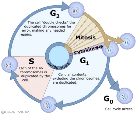
- G1 phase. Metabolic changes prepare the cell for division. At a certain point - the restriction point - the cell is committed to division and moves into the S phase.
- S phase. DNA synthesis replicates the genetic material. Each chromosome now consists of two sister chromatids.
- G2 phase. Metabolic changes assemble the cytoplasmic materials necessary for mitosis and cytokinesis.
- M phase. A nuclear division (mitosis) followed by a cell division (cytokinesis).
- The period between mitotic divisions - that is, G1, S and G2 - is known as interphase.
Mitosis
Mitosis is a form of eukaryotic cell division that produces two daughter cells with the same genetic component as the parent cell. Chromosomes replicated during the S phase are divided in such a way as to ensure that each daughter cell receives a copy of every chromosome. In actively dividing animal cells, the whole process takes about one hour.
The replicated chromosomes are attached to a 'mitotic apparatus' that aligns them and then separates the sister chromatids to produce an even partitioning of the genetic material. This separation of the genetic material in a mitotic nuclear division (or karyokinesis) is followed by a separation of the cell cytoplasm in a cellular division (or cytokinesis) to produce two daughter cells.
In some single-celled organisms mitosis forms the basis of asexual reproduction. In diploid multicellular organisms sexual reproduction involves the fusion of two haploid gametes to produce a diploid zygote. Mitotic divisions of the zygote and daughter cells are then responsible for the subsequent growth and development of the organism. In the adult organism, mitosis plays a role in cell replacement, wound healing and tumour formation.
Mitosis, although a continuous process, is conventionally divided into five stages: prophase, prometaphase, metaphase, anaphase and telophase.
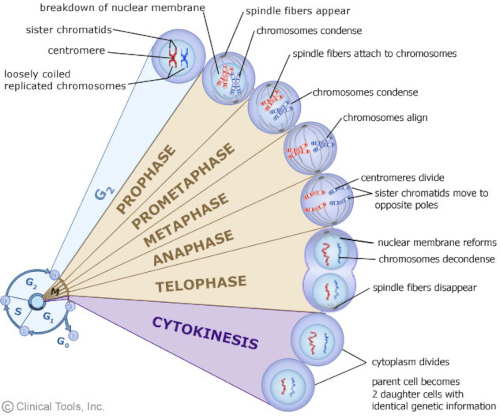
The phases of mitosis
Prophase
Prophase occupies over half of mitosis. The nuclear membrane breaks down to form a number of small vesicles and the nucleolus disintegrates. A structure known as the centrosome duplicates itself to form two daughter centrosomes that migrate to opposite ends of the cell. The centrosomes organise the production of microtubules that form the spindle fibres that constitute the mitotic spindle. The chromosomes condense into compact structures. Each replicated chromosome can now be seen to consist of two identical chromatids (or sister chromatids) held together by a structure known as the centromere.
Prometaphase
The chromosomes, led by their centromeres, migrate to the equatorial plane in the mid-line of the cell - at right-angles to the axis formed by the centrosomes. This region of the mitotic spindle is known as the metaphase plate. The spindle fibres bind to a structure associated with the centromere of each chromosome called a kinetochore. Individual spindle fibres bind to a kinetochore structure on each side of the centromere. The chromosomes continue to condense.
Metaphase
The chromosomes align themselves along the metaphase plate of the spindle apparatus.
Anaphase
The shortest stage of mitosis. The centromeres divide, and the sister chromatids of each chromosome are pulled apart - or 'disjoin' - and move to the opposite ends of the cell, pulled by spindle fibres attached to the kinetochore regions. The separated sister chromatids are now referred to as daughter chromosomes. (It is the alignment and separation in metaphase and anaphase that is important in ensuring that each daughter cell receives a copy of every chromosome.)
Telophase
The final stage of mitosis, and a reversal of many of the processes observed during prophase. The nuclear membrane reforms around the chromosomes grouped at either pole of the cell, the chromosomes uncoil and become diffuse, and the spindle fibres disappear.
Cytokinesis
The final cellular division to form two new cells. In plants a cell plate forms along the line of the metaphase plate; in animals there is a constriction of the cytoplasm. The cell then enters interphase - the interval between mitotic divisions.
Meiosis
Meiosis is the form of eukaryotic cell division that produces haploid sex cells or gametes (which contain a single copy of each chromosome) from diploid cells (which contain two copies of each chromosome). The process takes the form of one DNA replication followed by two successive nuclear and cellular divisions (Meiosis I and Meiosis II). As in mitosis, meiosis is preceded by a process of DNA replication that converts each chromosome into two sister chromatids.
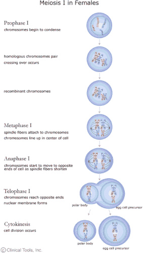
Meiosis I
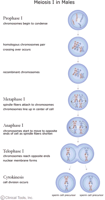
Meiosis I separates the pairs of homologous chromosomes.
In Meiosis I a special cell division reduces the cell from diploid to haploid.
Prophase I
The homologous chromosomes pair and exchange DNA to form recombinant chromosomes. Prophase I is divided into five phases:
- Leptotene: chromosomes start to condense.
- Zygotene: homologous chromosomes become closely associated (synapsis) to form pairs of chromosomes (bivalents) consisting of four chromatids (tetrads).
- Pachytene: crossing over between pairs of homologous chromosomes to form chiasmata (sing. chiasma).
- Diplotene: homologous chromosomes start to separate but remain attached by chiasmata.
- Diakinesis: homologous chromosomes continue to separate, and chiasmata move to the ends of the chromosomes.
Prometaphase I
Spindle apparatus formed, and chromosomes attached to spindle fibres by kinetochores.
Metaphase I
Homologous pairs of chromosomes (bivalents) arranged as a double row along the metaphase plate. The arrangement of the paired chromosomes with respect to the poles of the spindle apparatus is random along the metaphase plate. (This is a source of genetic variation through random assortment, as the paternal and maternal chromosomes in a homologous pair are similar but not identical. The number of possible arrangements is 2n, where n is the number of chromosomes in a haploid set. Human beings have 23 different chromosomes, so the number of possible combinations is 223, which is over 8 million.)
Anaphase I
The homologous chromosomes in each bivalent are separated and move to the opposite poles of the cell
Telophase I
The chromosomes become diffuse and the nuclear membrane reforms.
Cytokinesis
The final cellular division to form two new cells, followed by Meiosis II. Meiosis I is a reduction division: the original diploid cell had two copies of each chromosome; the newly formed haploid cells have one copy of each chromosome.
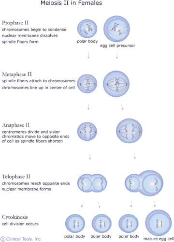
Meiosis II
Meiosis II separates each chromosome into two chromatids.
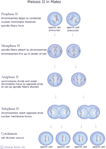
The events of Meiosis II are analogous to those of a mitotic division, although the number of chromosomes involved has been halved.
Meiosis generates genetic diversity through:
- the exchange of genetic material between homologous chromosomes during Meiosis I
- the random alignment of maternal and paternal chromosomes in Meiosis I
- the random alignment of the sister chromatids at Meiosis II
