People
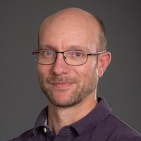
Prof Thomas Schalch
Professor of Molecular and Structural Biology

School/Department: Molecular Cell Biology Department of
Telephone: +44 (0)116 229 7139
Email: ts332@leicester.ac.uk
Profile
Professor Thomas Schalch has a keen interest in understanding how eukaryotic genomes work. He is an expert in structural biology and biochemistry with a focus on chromatin structure and function.
Research
The Schalch Lab specializes in exploring the structural and biochemical principles underpinning genome organization, with a particular focus on chromatin's role in gene regulation and its implications for cancer. Our research spans several key areas:
- Chromatin Structure and Gene Regulation: Investigating how chromatin fiber structure within the nucleus influences gene activity.
- Ubiquitination and Chromatin Signaling: Studying the role of ubiquitin in modulating chromatin dynamics, particularly how H2B ubiquitination prevents heterochromatin spread over active genes.
- Histone Modification and Heterochromatin Formation: Exploring the regulation of the Clr4 methyltransferase in heterochromatin assembly and gene silencing.
Our methodologies include X-ray crystallography, cryo-EM, genetic, and biochemical experiments, primarily using the fission yeast S. pombe as a model. We aim to elucidate the three-dimensional organization of chromatin and its impact on genome function, contributing to a deeper understanding of cellular mechanisms and potential therapeutic strategies.
Publications
Stirpe A. Guidotti N. Northall S. Kilic S. Hainard A. Vadas O. Fierz B. Schalch T. (preprint). SUV39 SET domains mediate crosstalk of heterochromatic histone marks. bioRxiv 2020.06.30.177071 Open Access Preprint
Leopold K. Stirpe A. and Schalch T. (2019). Transcriptional gene silencing requires dedicated interaction between HP1 protein Chp2 and chromatin remodeler Mit1. Genes & Development 33 565-577 Open Access Article
Ekundayo B. Richmond T.J. and Schalch T. (2017). Capturing structural heterogeneity in chromatin fibers. Journal of Molecular Biology 429 3031-3042. free article doi UNIGE archives Job
G. Brugger C. Xu T. Lowe B.R. Pfister Y. Qu C. Shanker S. Baños Sanz J.I. Partridge J.F. and Schalch T. (2016). SHREC Silences Heterochromatin via Distinct Remodeling and Deacetylation Modules. Molecular Cell 62 207-221. Article
Kuscu C. Zaratiegui M. Kim H.S. Wah D.A. Martienssen R.A. Schalch T. and Joshua-Tor L.(2014). CRL4-like Clr4 Complex in Schizosaccharomyces Pombe Depends on an Exposed Surface of Dos1 for Heterochromatin Silencing. PNAS 111 1795-1800. Article
Schalch T. Job G. Shanker S Partridge J. F. Joshua-Tor L. (2011). The Chp1-Tas3 core is a multifunctional platform critical for gene silencing by RITS. Nature Structural and Molecular Biology 18 1351-7. Article
Schalch T. Job G. Noffsinger V. J. Shanker S. Kuscu C. Joshua-Tor L. and Partridge J. F. (2009). High-affinity binding of Chp1 chromodomain to K9 methylated histone H3 is required to establish centromeric heterochromatin. Molecular Cell 34 36-46. Article
Schalch T. Duda S. Sargent D. F. and Richmond T. J. (2005). X-ray structure of a tetranucleosome and its implications for the chromatin fibre. Nature 436 138-41. Article
Dorigo B. Schalch T. Kulangara A. Duda S. Schroeder R. R. and Richmond T. J. (2004). Nucleosome arrays reveal the two-start organization of the chromatin fiber. Science 306 1571-3. Article
Dorigo B. Schalch T. Bystricky K. and Richmond T. J. (2003). Chromatin fiber folding: requirement for the histone H4 N-terminal tail. Journal of Molecular Biology 327 85-96. Article
Supervision
Thomas has successfully supervised 9 PhD students and many undergraduate and Masters students.
We are always looking for talented and motivated students to join our efforts. If you are interested please don't hesitate to get in touch with an email explaining your motivation and interests.
Teaching
Thomas Schalch values teaching highly and strives to deliver engaging and meaningful lectures and modules. He currently teaches on undergraduate biochemistry, molecular biology and gene expression modules.
Qualifications
- Ph.D. in Natural Sciences, ETH Zurich, Switzerland
- Diploma in Biology, ETH Zurich, Switzerland