New nanomaterial could transform how we visualise fingerprints
Innovative nanomaterials have the potential to revolutionise forensic science, particularly in the detection of latent (non-visible) fingermarks, following research conducted at Diamond’s labSAXS instrument (P38) and involving a University of Leicester scientist.
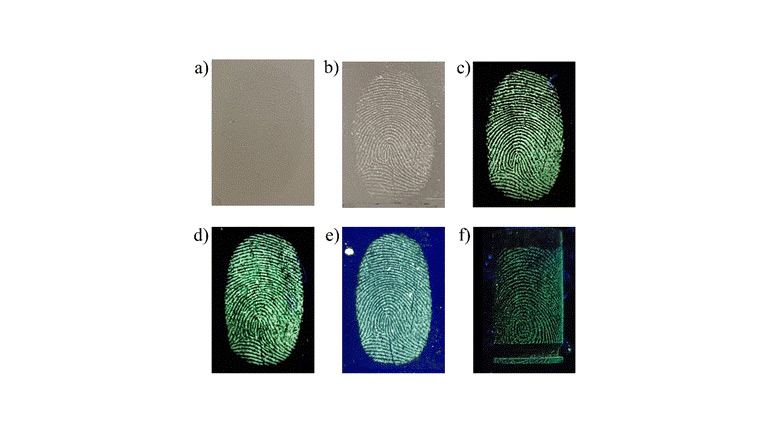
Researchers created a fluorescent nanoparticle using a combination of materials (MCM-41, chitosan and dansylglycine) to examine latent fingermarks. These nanoparticles have special properties that make them adhere well to fingerprint residues, even old ones. The nanoparticles work on various surfaces, including metal, plastic, glass and complex objects such as polymer banknotes. They have the potential to be used directly at crime scenes without lab facilities, which is a significant advantage over some previous reagents. They produce high-quality fingerprint images, with the vast majority of those tested meeting the UK Home Office standards for a successful identification. This new method captures the finer details of a fingermark, making it easier to identify individuals and is expected greatly to aid in forensic investigations.
The research was published in a Royal Society of Chemistry paper, highlighting that the new nanomaterial has proven to be a versatile and effective tool for visualising fingermark evidence. Small angle X-ray scattering (SAXS) techniques at Diamond provided useful data to validate these results.
The research team includes scientists from the Technical and Scientific Section of Alagoas, Federal Police, Brazil; the National Institute of Criminalistics of the Federal Police, Brazil; the University of Leicester’s School of Chemistry; the Federal University of Alagoas, Brazil; and the UK’s national synchrotron, Diamond Light Source.
Ridge patterns on fingertips remain unchanged during and beyond a person’s life. They provide the primary method of personal identification in criminal investigations. When an object’s surface is touched by a finger, sweat and oily substances are transferred and deposited onto the surface, resulting in the formation of a mark. Most fingermarks are invisible to the naked eye and are referred to as latent fingermarks.
The international collaboration of researchers developed the new nanostructured hybrid material, MCM-41@chitosan@dansylglycine, to visualise latent fingermarks. This material combines mesoporous silica nanoparticles with a fluorescent dye (dansylglycine) and chitosan, a polysaccharide derived from the exoskeletons of shrimps, crabs and lobsters.
Latent fingermarks require physicochemical development techniques to enhance their visibility and make them interpretable for forensic purposes. Traditional methods for developing fingerprints include optical, physical, and chemical processes that involve interaction between the developing agent (often a coloured or fluorescent reagent) and the fingermark residue. These methods have limitations in recovering high-quality results in certain conditions.
Recently, new methods using mass spectrometry, spectroscopy, electrochemistry, and nanoparticles have improved the development of latent fingermarks. These techniques offer better contrast, sensitivity, and selectivity, with low toxicity. The ability to adjust nanomaterial properties further enhances the detection of both fresh and aged fingermarks.
Mesoporous silica nanoparticles (MSNs) have attracted significant interest since the discovery of the M41S family of molecular sieves, which encompasses MCM-41, MCM-48, and SBA-15. These nanoparticles are characterised by their controlled particle size, porosity, high specific surface area, chemical stability, and ease of surface functionalisation.
Copyright of Diamond Light Source Ltd
Profa. Adriana Ribeiro, Federal University of Alagoas comments: “There are few studies employing chitosan for detection and enhancement of latent fingerprints and, to the best of our knowledge, no reports of the use of hierarchically structured MSNs modified with chitosan (MSN@Ch) for such applications – which was our strategy in this research. We exploited the MCM’s desirable characteristics – notably high surface area and surface modification – for the case of MCM-41 to enhance the interaction between the development reagent and fingerprint residue.”
The team added dansyl fluorophores which exhibit intense absorption bands in the near UV region and emit strong fluorescence in the visible spectrum with high emission quantum yields.
Professor of Physical Chemistry, Robert Hillman, University of Leicester concludes: “The overarching aim of this study was to create a versatile and effective latent fingermark visualisation material based on MSNs, chitosan and dansyl derivatives. These nanoparticles were applied as latent fingermark developers for marks on surfaces of diverse chemical composition, topography, optical characteristics and spatially variant nature, typical of forensically challenging evidence. For quality assessment of the enhanced fingermarks, we analysed the developed images using the UK Home Office scale, forensic protocols and, in terms of their constituent features, (minutiae), specialist forensic software. Across a substantive collection of marks deposited on chemically diverse surfaces and subject to complex environmental and temporal histories, the overwhelming majority of the enhanced images presented sufficient minutiae for comparison with model dactyloscopy images.”
Diamond Light Source CEO Prof. Gianluigi Botton adds: “It is pleasing to see that Diamond’s unique analytical tools once again have delivered outstanding science. Our network of international users is key to making sure our science delivers results. This advance in nanomaterials could be a step change in how forensics may be applied in the future.”