People
Professor Peter Moody
Professor of Structural Biology
School/Department: Molecular Cell Biology, Department of
Telephone: +44 (0)116 229 7097
Email: pcem1@leicester.ac.uk
Profile
I am Professor of Structural Biology. I read Biochemistry at York with a final year project using X-ray crystallography to determine the structure of a small bacterial ribonuclease. I continued using X-ray crystallography for my PhD in the Physics Department at Imperial College London with studies of the mechanism of the glycolytic enzyme Glyceraldehyde 3-Phosphate Dehydrogenase. After my PhD I continued to use protein crystallography to investigate enzyme mechanisms at Harvard, Imperial College and at York. I took a post at Leicester in 1995 where I work on the structures and mechanisms of redox and other enzymes using X-ray crystallography to determine structures and neutron crystallography in order see the positions of hydrogen atoms. We are currently developing ways to use electron diffraction in time-resolved studies.
Research
Enzyme structure and function
 We try to unravel enzyme mechanisms and investigate the way biological molecules recognise both smaller molecules and each other. Our primary technique is crystallography, X-ray crystallography is a very powerful technique that gives the three-dimensional structures of molecules, but the positions of hydrogen atoms are normally invisible. By using neutrons whose wavelength is similar to that of X-rays we are also able to determine the positions of these hydrogens. Together, neutron and X-ray crystallography allow us get detailed pictures of the interactions of biomolecules. Neutron crystallography has an additional advantage in that it avoids the ionising effects of X-irradiation. We use the nuclear reactors at the Institut Laue Langevan in Grenoble and MLZ Munich and the spallation source at Oak Ridge National Laboratories in Tennessee for neutrons and collaborate with the scientists at these facilities.
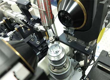
Because enzyme reactions are dynamic, we need ways to trap the intermediates in our crystals, and in order to follow the reactions we need to find ways to monitor them. In solution this is often done with spectrophotometry. We have a system for single crystal spectrophotometry that allows us to test if our methods of catching the various steps in the reaction have worked. We can then trap these intermediates in the crystal at low temperatures. Because we work with redox sensitive enzymes some of the work is done in our anaerobic glove box, this also houses a crystallisation robot.
Our work on heme peroxidases is in collaboration with Emma Raven and Hanna Kwon, our studies on molecular recognition in the complement system have been done with Russell Wallis. We also work on the enzymes in key metabolic pathways in pathogens, for example gluconeogenesis in enteric pathogens.
Publications
Selected publications:
Kwon et al. (2020) Visualizing the protons in a metalloenzyme electron proton transfer pathway. Proc. Natl. Acad Sci. (USA)117(12):6484-6490.
Kwon et al (2020) Heme peroxidase - Trapping intermediates by cryo neutron crystallography Methods in Enzymology Vol 634 (Ed. P C E Moody) https://doi.org/10.1016/bs.mie.2020.01.010
Moody PCE, Raven EL The Nature and Reactivity of Ferryl Heme in Compounds I and II Acc. Chem Res. 51(2):427-435
Kwon H, et al. (2017) 'Combining X-ray and neutron crystallography with spectroscopy.' Acta Crystallogr D Struct Bio, vol. 73. Pp. 141-147.
Kwon H, et al. (2016) 'Direct visualization of a Fe(IV)-OH intermediate in a heme enzyme.' Nat Commun, vol. 7. P. 13445.
Casadei CM, et al. (2014) 'Heme enzymes. Neutron cryocrystallography captures the protonation state of ferryl heme in a peroxidase.' Science, vol. 345. Pp. 193-197.
For full list of publications please see Peter's Google Scholar profile
Supervision
Teaching
- BS3070 Structural Biology
- BS7102 Proteins