Archaeology and Ancient History
Advanced Microanalysis Laboratory
The Advanced Microanalysis Laboratory houses a suite of instruments allowing for the analysis and identification of inorganic and organic materials at an atomic and molecular level. Analysis can include materials and artefacts and any kind of object made from inorganics such as stone (tools, masonry, etc.), ochre, pottery, ceramics, glass, metal and includes organic materials such as plant matter (archaeobotany), resins, adhesives, paper, vellum (illuminated manuscripts), fine art (paintings, drawings, print, etc.) organic residues etc. As well as providing excellent facilities for research, the suite offers students access to state-of-the-art instruments, ensuring our students graduate with a competitive edge and have a solid platform for moving into highly skilled employment.
Facilities in this laboratory include a brand-new Scanning Electron Microscope (SEM) equipped with Energy Dispersive X-ray Spectroscopy (EDS) and X-ray Fluorescence (XRF), Raman Spectrometer, Benchtop µXRF and Handheld pXRF. The laboratory also houses a Carl Zeiss AxioScan for high-resolution scanning of thin-sections.
Scanning Electron Microscope (SEM)
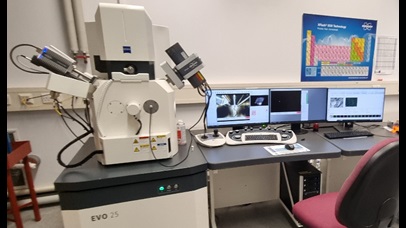 The SEM is a Carl Zeiss Evo 25,equipped with Bruker XFlash (EDS) and XTrace (XRF) detectors. The SEM has a large chamber size allowing for the analysis of large objects such as skulls, fossils, and large artefacts.
The EVO is a conventional SEM, often seen as a workhorse instrument allowing flexibility for sample analysis. It can use either a Tungsten or Lanthanum Hexaboride (LaB6) filaments, allowing for ~3nm resolution at 30kV. The SEM can analyse samples in high vacuum (conductive samples) and variable pressure (non-conductive samples). The SEM is equipped with Secondary Electron (SE1 and C2D) detectors allowing topographical information to obtained alongside a Backscattered Electron (BSE) detector which gives compositional information. Elemental analysis can be acquired from EDS and XRF detectors, this can be either as a compositional elemental map or as quantitative point analysis. The flexibility of both EDS and XRF detectors allows beam sensitive samples to be analysed using XRF as opposed to the electron beam, this penetrates into the sample further and can reveal trace elements. The EDS detector allows for lighter elements to be detected (Boron upwards) and gives more surface information.
The SEM is equipped with Zen Connect, allowing a correlative workflow between LM’s, SEM, FEG SEM (Field Emission SEM), FIB SEM (Focussed Ion Beam SEM) and XCT (Crystal CT) across campus. This enables the user to be able to look at the same location on a sample under any microscope, as long as the sample preparation is compatible.
M4 Tornado
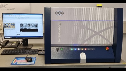 The Bruker M4 Tornado is a benchtop µXRF, allowing for samples to be imaged and examined in further detail with elemental mapping and point analysis. Data can be both qualitative and quantitative. Data from the M4 Tornado is compatible with the data acquired from the EDS and XRF detectors on the SEM, allowing for detailed sample analysis.
Axio Scan
The Carl Zeiss Axio Scan slide scanner allows automated digitisation of thin section slides, these can be biological, geological and petrography.The Axio Scan allows high speed digitisation and high-resolution image quality. The slide scanner can offer high throughput of samples with automated scanning of up to 100 slides in a single run. The system is equipped with fluorescence, brightfield, and polarisation capabilities.
Portable X-Ray Fluorescence (pXRF)
The Olympus VANTA portable X-Ray Fluorescence (pXRF) analyser (Figure 5) is a handheld energy-dispersive X-Ray Fluorescence spectrometer capable of delivering fast and precise identification and quantification of elements from Mg to U. The pXRF can be used for materials analysis on artefacts and soils both in the laboratory and in the field.
Raman Spectrometer
 The Raman spectrometer is complementary to established imaging techniques, as it offers chemical information as opposed to elemental information. Researchers will gain information regarding the chemical bonds within their sample, offering the ability to analyse the chemical composition of a sample at a molecular level. It can determine the composition pigment/ink samples, trace and residual analysis.